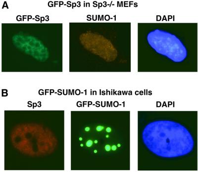
Fig. 6. Subcellular localization of Sp3 and SUMO-1 in MEFs and Ishikawa cells. (A) Sp3–/– MEFs were transfected with 1 µg of an expression construct for GFP–Sp3. The intracellular distribution of GFP–Sp3 was detected by intrinsic green fluorescence of the GFP tag. Endogenous SUMO-1 localization was detected with a rabbit anti-SUMO-1 antibody and a CY3-conjugated secondary antibody. (B) Ishikawa cells were transfected with 1 µg of an expression construct for GFP–SUMO-1. Visualization was by the intrinsic green fluorescence of the GFP moiety. Endogenous Sp3 localization was detected with a rabbit anti-Sp3 antibody and a CY3-conjugated secondary antibody.

An official website of the United States government
Here's how you know
Official websites use .gov
A
.gov website belongs to an official
government organization in the United States.
Secure .gov websites use HTTPS
A lock (
) or https:// means you've safely
connected to the .gov website. Share sensitive
information only on official, secure websites.
