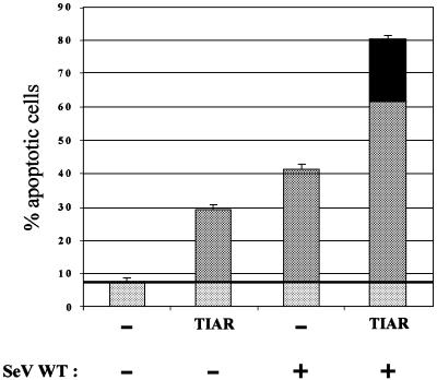
Fig. 8. Synergy of TIAR overexpression and SeV infection in inducing apoptosis. Duplicate cultures of HeLa cells were transfected with 1 µg of pEBS plasmids expressing TIAR or empty pEBS (as indicated), along with 0.2 µg of pEBS–GFP to follow transfection efficiency, with fuGENE (Roche). The cultures were then infected 24 h later with 10 p.f.u./cell of SeV-wt, or mock infected. All cultures were harvested at 48 h.p.i., and the percentage of apoptotic cells was determined by annexin V staining and FACS. The horizontal line indicates the background level of apoptosis, i.e. independent of TIAR expression and SeV infection, which is subtracted from the other bars to determine the level of synergy. The synergy between TIAR overexpression and SeV infection is indicated by the black segment at the top of the bar.

An official website of the United States government
Here's how you know
Official websites use .gov
A
.gov website belongs to an official
government organization in the United States.
Secure .gov websites use HTTPS
A lock (
) or https:// means you've safely
connected to the .gov website. Share sensitive
information only on official, secure websites.
