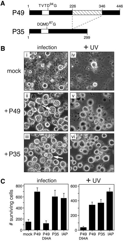
Fig. 1. P49 blocks apoptosis induced by diverse signals. (A) P49 and P35 structure. Amino acid similarity between P49 and P35 is colinear with the exception of a 120 residue insert (crosshatched) within P49. The caspase cleavage site within the predicted RSL (open) is indicated. (B) Virus- and UV radiation-induced apoptosis. SF21 cells were mock-transfected or transfected with plasmids encoding P49 or P35 and subsequently infected with apoptosis-inducing virus vΔp35 or irradiated with UV-B (125 mJ/cm2). Photographs (magnification, 100×) were taken 48 or 24 h after infection (i–iii) or UV irradiation (iv–vi), respectively. Arrows depict occluded virus particles in non-apoptotic cells. A representative experiment is shown. (C) Cell survival. SF21 cells transfected with plasmids encoding wild-type P49, D94A-mutated P49, P35 or Op-IAP were infected or UV-irradiated as described in (B). Survival was quantified by computer-aided microscopy and is reported as the average number of surviving, non-apoptotic cells ± standard deviation.

An official website of the United States government
Here's how you know
Official websites use .gov
A
.gov website belongs to an official
government organization in the United States.
Secure .gov websites use HTTPS
A lock (
) or https:// means you've safely
connected to the .gov website. Share sensitive
information only on official, secure websites.
