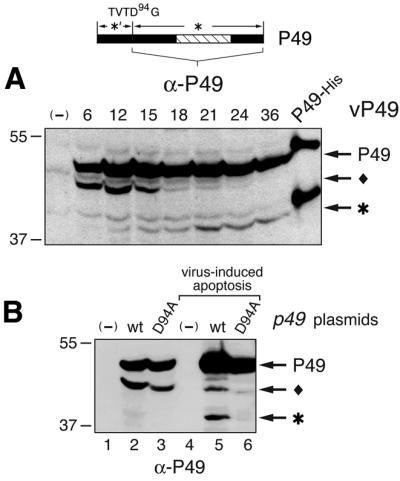
Fig. 3. In vivo P49 cleavage. (A) Time course. SF21 cell lysates prepared at the indicated hours after infection with vP49 were subjected to immunoblot analysis using α-P49, which recognized full-length P49 and the 40 kDa cleavage fragment (top), but not the N-terminal 9 kDa fragment. Due to the absence of His tags, in vivo P49 and its 40 kDa cleavage fragment (*) are smaller than P49-His6, which was partially cleaved by caspase (right lane). Another P49-related protein (filled diamond) is indicated. (B) Asp94-dependent P49 cleavage. SF21 cells were mock-transfected (–) or transfected with plasmid encoding wild-type (wt) or D94A-mutated P49. After mock infection (lanes 1–3) or infection with p35– vΔp35 to induce apoptosis (lanes 4–6), cell lysates (5 × 10⁵ cell equiv) were prepared and subjected to α-P49 immunoblot analysis.
