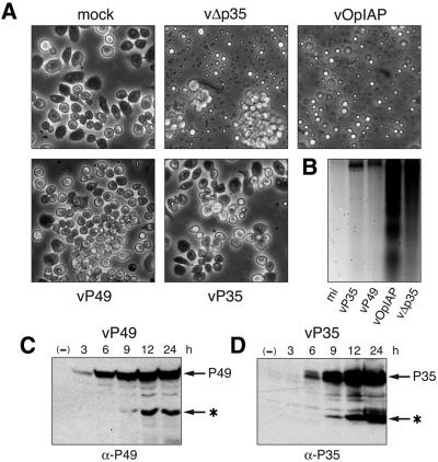
Fig. 8. P49 blocks baculovirus-induced apoptosis in Drosophila DL-1 cells. (A) Virus-induced apoptosis. Drosophila Line-1 (DL-1) cells were photographed 24 h after mock infection (mock) or inoculation with p35– vΔp35, Op-iap+ vOp-IAP, p49+ vP49 or wild-type p35+ virus (vP35). Magnification 100×. (B) DNA fragmentation. Low molecular weight DNA was selectively extracted from equivalent numbers of DL-1 cells and associated apoptotic bodies. Only fragmented DNA was visualized by agarose gel electrophoresis since intact cellular DNA was excluded. (C and D) P49 synthesis and cleavage. DL-1 lysates (3 × 10⁶ cell equiv) prepared at the times indicated after inoculation with vP49 or vP35 were subjected to immunoblot analysis with α-P49 (C) or α-P35 (D). Full-length and C-terminal cleavage (*) proteins are indicated.

An official website of the United States government
Here's how you know
Official websites use .gov
A
.gov website belongs to an official
government organization in the United States.
Secure .gov websites use HTTPS
A lock (
) or https:// means you've safely
connected to the .gov website. Share sensitive
information only on official, secure websites.
