
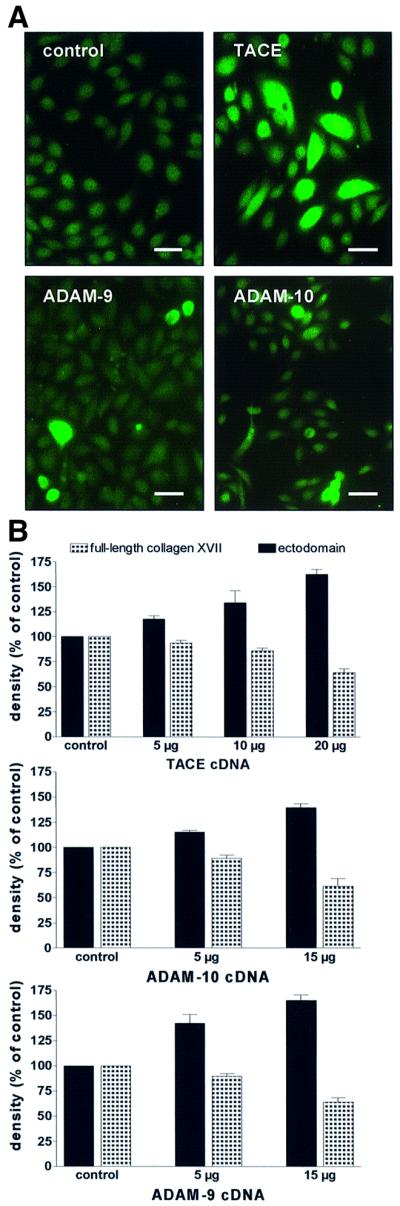
Fig. 5. Transient transfections of HaCaT cells with cDNAs for TACE, ADAM-9 and ADAM-10. (A) Immunofluorescence staining of transfected cells. Cells transfected with empty vector (control) remained negative, but ADAM-transfected cells showed strong positive signals. Scale bar: 100 µm. (B) HaCaT cells were transfected with different concentrations of cDNA for TACE (upper panel), ADAM-10 (middle panel) and ADAM-9 (lower panel) and shedding of collagen XVII was assessed with immunoblotting with a mixture of antibodies Endo-2, NC16A and Ecto-1. Densitometric analysis of the signals (expressed as a percentage of the control ± SD; n = 3) showed a dose-dependent increase of ectodomain shedding, with a concomitant decrease of full-length collagen XVII.