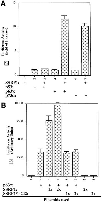
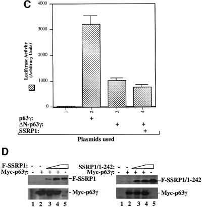
Fig. 1. SSRP1 enhances the transcriptional activity of p63γ and p73α, but not of p53 and ΔN-p63γ. (A) SSRP1 stimulates the transcriptional activity of p63 and p73, but not p53. H1299 cells were transfected with plasmids encoding p53 (50 ng), p63γ (50 ng) or p73α (50 ng) alone or with SSRP1 (750 ng) as indicated, along with reporters as described in Materials and methods. At 48 h post-transfection, cells were harvested for luciferase and β-gal assays. Luciferase activity was normalized by the internal β-gal activity and expressed in fold increase (each column represents the mean activity from three independent assays, and bars show standard deviation). (B) SSRP1, but not its N-terminal fragment, stimulates the p63γ-dependent luciferase activity. H1299 cells were transfected with pCDNA-Myc-p63γ (50 ng), pCDNA3-Flag-SSRP1 (1× 250 ng and 2× 500 ng) and pCDNA3-Flag-SSRP1/1–242 (500 ng) as indicated. Luciferase and β-gal assays were conducted as described above. Luciferase activity was expressed in arbitrary units here. (C) SSRP1 does not stimulate the ΔN-p63γ-dependent luciferase activity. The same transfection was carried out as that described above, except that pCDNA3-Myc-ΔN-p63γ (50 ng) was also used here in comparison with p63γ. A 50 ng aliquot of pCDNA3-Myc-p63γ and 500 ng of pCDNA3-Flag-SSRP1 were used in this experiment. The results shown in (A–C) were reproduced using p53 null H1299 cells and MEFs. (D) Western blot analysis of expression of exogenous p63γ and SSRP1. H1299 cells were transfected as described in the above panels, except that triplicate plates were used in each lane for detection of proteins. A 150 µg aliquot of proteins was loaded directly on an SDS gel. Flag-SSRP1 (F-SSRP1), Flag-SSRP1/1–242 (F-SSRP1/1–242) or Myc-p63γ was detected by western blot analyses using monoclonal anti-Flag or anti-Myc antibodies.

An official website of the United States government
Here's how you know
Official websites use .gov
A
.gov website belongs to an official
government organization in the United States.
Secure .gov websites use HTTPS
A lock (
) or https:// means you've safely
connected to the .gov website. Share sensitive
information only on official, secure websites.

