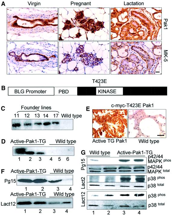
Fig. 3. Generation of active Pak1-TG mice. (A) Expression of Pak1 during various stages of mammary gland development as revealed by immunohistochemistry. The lower panels show myoepithelial cells identified by the mouse keratin-5 marker (MK-5). (B) Structure of the T423E Pak1 transgene. c-myc-tagged T423E-mutated Pak1 was fused to the ovine BLG promoter. PBD, p21-binding domain. (C) Identification of the TG founder mice by Southern blotting. Tail DNA (5 µg) was digested with BamHI, resolved on 0.7% agarose gel and probed with a Pak1 cDNA probe. The transgene band is 2.2 kb. (D) Expression of transgene as determined by RT–PCR followed by Southern blotting. Three Pak1-TG mice on day 12 of lactation and two wild-type mice were analyzed on the same day of lactation. (E) Expression of transgene on day 12 of lactating mammary glands as revealed by immunohistochemistry using anti-c-myc-tag antibody. An age-matched wild-type mammary gland was used as a control. (F) Pak1 kinase activity analysis. Protein lysates from the indicated days of pregnancy or lactation were immunoprecipitated with Pak1 antibody and then in vitro kinase assay was performed using myelin basic protein (MBP) as a substrate. (G) Western blotting of mammary lysates showed an increased level of phosphorylated p38MAPK and MAPK in the Pak1-TG mice, with an unchanged level of total p38 and MAPK. Bar = 40 µm.

An official website of the United States government
Here's how you know
Official websites use .gov
A
.gov website belongs to an official
government organization in the United States.
Secure .gov websites use HTTPS
A lock (
) or https:// means you've safely
connected to the .gov website. Share sensitive
information only on official, secure websites.
