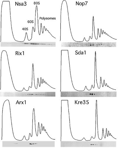
Fig. 1. Reverse-tagged protein baits of the Nug1-containing 60S pre- ribosome are associated with pre-ribosomal particles of different sizes. The sedimentation behavior of the indicated TAP-tagged proteins was analyzed on sucrose density gradients, and ribosomal fractions (40S, 60S, 80S and polysomes) were determined by OD254 measurement of the gradient fractions (upper graph). Western blot analysis of these gradient fractions using anti-ProtA antibodies reveals the position of the indicated TAP-tagged baits (lower panel). Note that some baits (e.g. Nsa3) exhibit a broad distribution on the sucrose gradient, whereas other baits (e.g. Kre35) exhibit a distinct peak at ∼60S (see text).

An official website of the United States government
Here's how you know
Official websites use .gov
A
.gov website belongs to an official
government organization in the United States.
Secure .gov websites use HTTPS
A lock (
) or https:// means you've safely
connected to the .gov website. Share sensitive
information only on official, secure websites.
