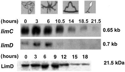
Fig. 2. Presence of limC and limD mRNAs and protein during development. The same amount (30 µg) of total RNA isolated from cells developed on phosphate-buffered agar plates at different hours of development as indicated was loaded in each lane. Hybridization was with 32P-labeled limC or limD cDNA. mRNA sizes are given in kb. For detection of LimD protein, total cellular extracts from AX2 cells (4 × 10⁵ cells per lane) harvested at different time points were separated by SDS–PAGE (15% acrylamide). The resulting western blot was probed with LimD-specific monoclonal antibody K4-353-6.

An official website of the United States government
Here's how you know
Official websites use .gov
A
.gov website belongs to an official
government organization in the United States.
Secure .gov websites use HTTPS
A lock (
) or https:// means you've safely
connected to the .gov website. Share sensitive
information only on official, secure websites.
