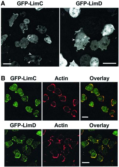
Fig. 3. Distribution of GFP–LimC and GFP–LimD fusion proteins. (A) Both the fusion proteins accumulate to high levels at distinct areas of the cell cortex and are distributed throughout the cytoplasm. Occasional localization of both the fusion proteins in the nucleus was also observed (arrowheads). Bars, 10 µm. (B) GFP–LimC (upper panels) and GFP–LimD (lower panels) localization coincides with that of actin in the cell cortex. The cells were fixed with cold methanol and immunolabeled with anti-actin monoclonal antibody followed by Cy3-conjugated anti-mouse secondary antibody. Overlay images show the co-localization of GFP–LimC or GFP–LimD fluorescence with the actin staining in the cell cortex. Bars, 10 µm.

An official website of the United States government
Here's how you know
Official websites use .gov
A
.gov website belongs to an official
government organization in the United States.
Secure .gov websites use HTTPS
A lock (
) or https:// means you've safely
connected to the .gov website. Share sensitive
information only on official, secure websites.
