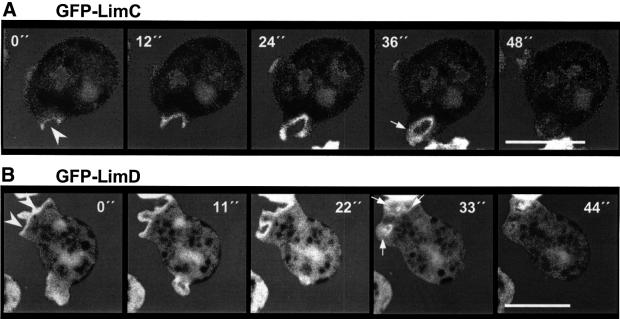
Fig. 4. GFP–LimC and GFP–LimD redistribution during fluid-phase endocytosis. Cells expressing GFP–LimC (A) or GFP–LimD (B) were allowed to adhere to glass coverslips and the supernatant was replaced by phosphate buffer containing 1 mg/ml TRITC–dextran. Confocal sections were taken at the times indicated. Arrowheads in the 0 s panels indicate the site of formation of pinocytic vesicles and arrows in subsequent images indicate newly formed pinosomes. Bars, 10 µm.

An official website of the United States government
Here's how you know
Official websites use .gov
A
.gov website belongs to an official
government organization in the United States.
Secure .gov websites use HTTPS
A lock (
) or https:// means you've safely
connected to the .gov website. Share sensitive
information only on official, secure websites.
