FIG. 3.
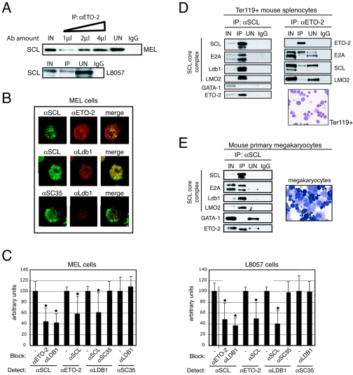
Validation of the SCL/ETO-2 interaction in cell lines and primary cells. (A) Anti-ETO-2 (αETO-2) antibodies (Ab) immunoprecipitate SCL from MEL (top panel) and L8057 (bottom panel) nuclear extracts. The triangle represents increasing amounts of αETO-2 antibodies. IgG, negative control. (B) Nuclear colocalization of SCL and ETO-2 in MEL cells was detected by immunofluorescence (upper panel, first row, merge image). Incubation with αSCL/αLdb-1 antibodies served as a positive control (second row). Incubation with αLdb-1/αSC35 antibodies served as a negative control (third row). (C) Colocalization is revealed by antibody blocking in MEL (left panel) and L8057 (right panel) nuclei. Anti-ETO-2 and anti-Ldb-1 antibodies prevent access of anti-SCL antibodies to the antigen and vice versa. Anti-SC35 antibodies have no blocking effect on anti-Ldb-1 (and vice versa). Antibodies used for blocking and detection are shown. *, P value <0.05. (D and E) Nuclear extracts prepared from Ter119+ mouse splenocytes (D) and primary megakaryocytes (E) were subjected to coimmunoprecipitation with anti-SCL or anti-ETO-2 (splenocytes only) antibodies. Immunoprecipitated proteins were detected by Western blot. Morphology of Ter119+ cells and megakaryocytes used for nuclear extract preparation was assessed by May-Grunwald-Giemsa staining. Ter119+ cells represent proerythroblasts to mature erythrocyte and enucleated stages. Primary megakaryocytes comprise immature megakaryocyte precursors (dark cytoplasm) and mature megakaryocytes (large cells with a granular cytoplasm and a polyploid nucleus).

