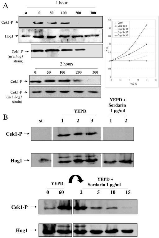
FIG. 7.
Growth-dependent activation of the Cek1 MAP kinase. (A) Stationary-phase cells of the wt strain (RM100) were diluted at an A600 of 0.1 in either YEPD or YEPD in the presence of Congo red at 50, 100, 200, and 300 μg/ml. Samples were collected after 1 and 2 h of growing at 37°C, and cells were processed for MAP kinase phosphorylation by Western blotting analysis. The increase in A600 is shown in the diagram at the right. Symbols are the same as described in the legend to Fig. 6. Results for the hog1 mutant under the same conditions are also shown. (B) An overnight culture of a wt strain (labeled st) was diluted in fresh YEPD to an A600 of 0.2 and incubated at 37°C, taking samples after 1, 2, and 3 h of growth in these conditions (shown in each lane; YEPD lanes 1, 2, and 3). A portion of the overnight culture was also transferred to a prewarmed flask with YEPD medium, where 1 μg/ml sordarin was added, and samples were taken after 1 and 2 h of incubation at 37°C and processed (labeled YEPD + sordarin, lanes 1 and 2). Lanes are arranged in this figure from a scanned Western blot (as shown by blank intermediate lines) to avoid incorporation of samples not related to this particular experiment. They correspond to the same experiment and were processed simultaneously in the same gel. (C) Similarly, stationary growing cells of a wt strain were diluted in fresh YEPD to an A600 of 0.2 and incubated at 37°C. After 1 h, 1 μg/ml sordarin was added. Samples were collected at the times indicated in each lane (in minutes). Symbols are same as described in the legend to Fig. 6.

