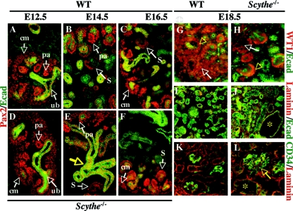
FIG. 3.
Analysis of Scythe−/− kidneys. (A to F) Immunostaining of Pax2 (red) and E-cadherin (Ecad) (green) with control (A, B, and C) and mutant (D, E, and F) embryos was done at E12.5, E14.5, and E16.5. At E12.5, the ureteric bud (ub) was surrounded by condensed mesenchyme (A and D), and ureteric bud branching and formation of pretubular aggregates (pa) could be seen for both WT and Scythe−/− embryos. Pax2 was expressed in condensed mesenchymal cells (cm), pretubular aggregates, and comma- and S-shaped bodies (S) in both WT and Scythe−/− embryos at E14.5 and E16.5, but nephrogenic differentiation was delayed in Scythe−/− embryos (B, C, E, and F). The structure of the epithelium (Ecad staining) showed branching abnormalities (E, yellow arrow) in Scythe−/− kidneys. (G to L) At E18.5, WT1 (red) and E-cadherin (green) expression was detected in the condensing blastema (arrows), the proximal part of S-shaped bodies, and the podocytes of functional glomeruli (arrowheads) for both WT and Scythe−/− kidney (G and H) but to a lesser extent in Scythe−/− kidney. Laminin (red) distribution in Scythe−/− kidneys at E18.5 showed a pattern similar to that of the WT, while E-cadherin staining (green) showed enlarged tubules (asterisk) in Scythe−/− kidneys (I and J). The endothelial marker CD34 (green) is expressed similarly in the WT and in Scythe−/−, although enlarged glomeruli (arrow) and tubules (asterisk) are seen with Scythe−/− kidney (K and L).

