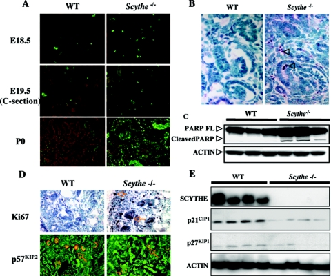
FIG. 5.
Increased apoptosis and cellular proliferation occurs in Scythe−/− kidneys. (A) TUNEL assays of kidney sections at E18.5, E19.5, and P0 showed increased apoptosis in Scythe−/− kidneys. C-section, cesarean section. (B) Staining with anti-cleaved caspase 3 (arrowheads) showed increased apoptosis in cortical mesenchyme from E18.5 Scythe−/− kidneys. (C) Proteins were isolated from three WT and three Scythe−/− E18.5 kidneys, and PARP cleavage was analyzed by Western blot. Actin protein levels were used as a loading control. FL, full length. (D) Ki67 and p57KIP2 immunohistochemistry showed increased proliferation in Scythe−/− kidneys at E18.5 compared to the WT control. (E) Western blot analysis of cyclin-dependent kinase inhibitors p21CIP1 and p27KIP1 from multiple individual WT and Scythe−/− E18.5 kidneys showed expression patterns consistent with increased proliferation. Scythe and actin protein levels were used as controls.

