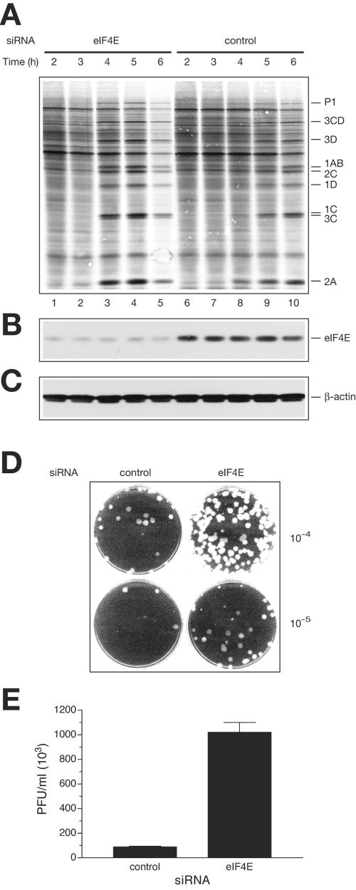
FIG. 4.
eIF4E knockdown stimulates translation and replication of EMCV in vivo. (A) Time course of protein synthesis in EMCV-infected cells. siRNA against eIF4E or a nonspecific siRNA (con- trol) was transfected into HeLa S3 cells. eIF4E knockdown or control cells were infected with EMCV, and protein synthesis was examined by pulse-labeling with [35S]methionine at the indicated time points. After labeling, polypeptides were resolved by SDS-PAGE and transferred to a PVDF membrane. The autoradiograph of the membrane is shown. The positions of the major EMCV-specific proteins are indicated on the right. (B) eIF4E levels in cells, as analyzed by Western blotting. The membrane from panel A was probed with anti-eIF4E, and signals were quantified as described in Materials and Methods. The average level of eIF4E depletion for lanes 1 to 5 (versus lanes 6 to 10) was 86%. (C) β-Actin detection by Western blotting (a loading control). (D) Plaque assays of the indicated dilutions of the lysates from control and eIF4E knockdown cells 4 h after infection. (E) EMCV yield, as affected by eIF4E siRNA treatment. EMCV-infected cells (eIF4E knockdown or control, unlabeled) were lysed at 4 h postinfection. Viral titer was measured as described in the legend to Fig. 2D.

