FIG.3.
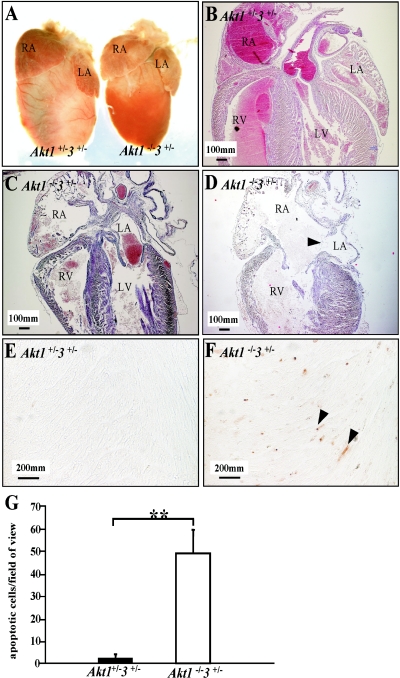
Structural defects in the heart from Akt1−/− Akt3+/− mice. (A) The hearts of Akt1−/− Akt3+/− mice are smaller than those of the controls, but the atria are as big as those of the controls. (B to D) Sagittal sections of the hearts. (B to D) HE staining of hearts from panel A. (C and D) Enlargement of the right side of Akt1−/− Akt3+/− hearts and apparent atria septum defects in Akt1−/− Akt3+/− hearts (P3). The wall of the right atrium of the Akt1−/− Akt3+/− heart is thin due to enlargement (C and D). The arrowhead in panel D indicates atria septum defects. (E and F) TUNEL assay. Arrowheads indicate apoptotic cells. (G) Quantification of apoptosis. Error bars indicate standard deviation. **, P < 0.01. RA, right atrium; RV, right ventricle; LA, left atrium. Magnification for panels B to D, ×40; for panels E and F, ×400.

