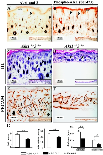
FIG.4.
Histology of Akt1−/− Akt3+/− skin. (A and B) Immunohistochemical staining of Akt1/Akt3 and phospho-Akt (Ser473) in control skin. (A) Akt1/Akt3 localization. The two proteins are mainly localized in the keratinocytes of the epidermis (Ep) and hair follicles (arrowheads). (B) Phospho-Akt (Ser473) in control skin. Localization of phospho-Akt is similar to that of Akt1/Akt3. (C and D) HE staining. Skin of Akt1−/− Akt3+/− mice is much thinner than that of control littermates and has fewer hair follicles (arrowheads). (E and F) PECAM (CD31) staining to display the vasculature. Akt1−/− Akt3+/− skin has fewer vessels than the control. Insets in all panels are lower magnification images showing a larger part of the skin. (G) Quantification of skin thickness and hair follicles. HE staining sections from three Akt1−/− Akt3+/− mice and three Akt1+/− Akt3+/− littermates (P3) were measured for thickness and hair follicle density. Error bars are standard deviation, and two asterisks indicate a significant difference between the two genotypes (P < 0.05). Magnification, ×200.

