FIG.5.
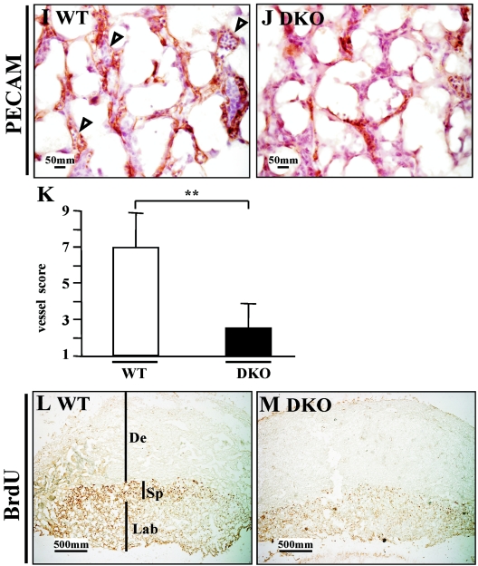
Analysis of the vascular system of DKO embryos. (A to H) Left panels are HE staining to show the morphology, and the right panels are PECAM staining to display vasculature. (A to D) Fine vessels (capillary) in the neuroepithelium of hindbrain. Arrowheads indicate the small vessels (capillary). Small vessels are abundant in the control and are evenly distributed, while these vessels are scarce in DKO embryos. (E to H) Branchial arches and their arteries. In the control, first, second, and third arch arteries can be seen (arrowheads), and small vessels are rich in mesenchymal tissue (F). However, the branchial arches of DKO mice are hypoplastic, with little mesenchymal tissue, and the large arch arteries cannot be distinguished (arrowhead). Small vessels are also scarce in DKO branchial arches. Abbreviations: NE, neuroepithelium; BA, branchial arch; WT, wild type. Magnification, ×100. (I to M) Histological analysis of DKO placenta. (I and J) E12.5 placentas. PECAM staining displays fetal vessels in the labyrinth. In the wild type (I), fetal vessels are well formed and filled with fetal nucleated erythroid cells (arrowheads), while in DKO mice (J) these vessels are rare, although the epithelial cells can be seen. (K) Quantification of fetal vessels in wild-type and DKO placenta. The double asterisks indicates a significant difference (P < 0.05). (L and M) E10.5 placentas. BrdU staining. DKO placenta shows a substantial reduction of proliferation, especially in the spongiotrophoblast layer (Sp). De, decidua; Lab, labyrinth. Magnification, ×40.


