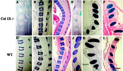
FIG. 2.
Histological and immunohistological analysis of newborn mice. Paraffin-embedded sagittal sections of newborn collagen IX knockout (A to F) and wild-type (G to L) mice were stained with antibodies to collagen IX (A, D, G, and J) or collagen II (B, E, H, and K) and with alcian blue (C, F, I, and L). Panels A to C and G to I show vertebral bodies, and panels D to F and J to L display ribs. All cartilaginous and cartilage primordial tissues were stained similarly for collagen II and with alcian blue regardless of genotype (B, C, E, and F; H, I, K, and L). However, collagen IX-deficient cartilage did not show any staining with antibodies to collagen IX (A and D). Bar, 550 μm.

