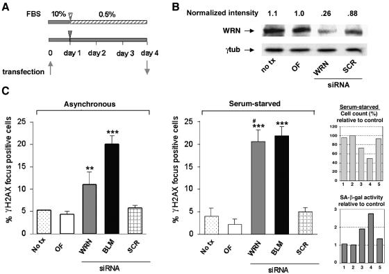
FIG. 4.
Preferential accumulation of DNA damage foci by acute withdrawal of WRN in serum-synchronized cells. (A) Primary WI-38 cells (PDL 26) at ∼30% confluence were transfected with the various siRNAs and either cultured in the presence of 10% serum (asynchronous) or synchronized with 0.5% serum for 72 h to put the cell into the G0 state, as shown by the experimental time line. (B) Quantitative immunoblot analysis (as described in the legend to Fig. 1) shows the extent of WRN depletion at day 4 posttransfection in serum-starved WI-38 cells. The concentration of the various siRNAs was 50 nM. (C) Quantitation of γH2AX foci. Histograms indicate the percentage of cells with foci (for criteria, see Materials and Methods) in primary asynchronous (left panel) or serum synchronized (right panel) WI-38 cells 4 days after various siRNA transfections. On average, more than 250 intact nuclei were screened per individual condition, and the data were derived from at least four independent biological experiments. Error bars represent the SEM. **, level of significance with a P value of <0.05; ***, level of significance with a P value of <0.005 (compared to the respective OF control was made by using Student t test). The “#” symbol indicates a statistically significant difference (P < 0.05) in γH2AX foci formation between serum-synchronized and asynchronous WRN depleted cells. The inset shows the cell number and SA-β-Gal expression in serum-synchronized cells after RNAi; the average of two independent experiments is shown. Columns: 1, no treatment; 2, OF, 3, WRN siRNA; 4, BLM siRNA; 5, SCR siRNA. OF, Oligofectamine.

