FIG. 7.
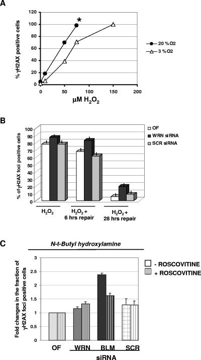
Increased sensitivity of WRN-depleted cells to different types of oxidative damage. (A) Sensitivity of normal fibroblasts, maintained in ambient (20%) or 3% oxygen, to form γH2AX foci in response to various doses of hydrogen peroxide treatment. Asynchronous WI-38 cells were cultured under 20% and 3% oxygen and treated for 45 min with the indicated doses of H2O2 in serum free-medium, followed by fixation and immunofluorescence analysis. The asterisk indicates a mild to moderate amount of cell death in 20% oxygen, which became predominant at a 150 μM H2O2 dose. (B) Prolonged presence of DNA damage foci induced by acute hydrogen peroxide treatment in WRN-depleted cells. WI-38 cells were cultured under 3% oxygen. At 96 h after siRNA transfection, the cells were treated for 45 min with 75 μM H2O2 in serum free-medium then either processed immediately for immunofluorescence analysis or, after medium change, allowed to recover at 3% oxygen for various amounts of time, as indicated. Bars indicate the percentage of γH2AX focus-positive cells. Error bars show the SEMs of three experiments. (C) The antioxidant NtBHA protects WRN-depleted cells to develop DNA damage foci at ambient oxygen. The cells were subcultured at 20% oxygen in the presence of 100 μM NtBHA for 2 weeks and then transfected with the various siRNAs and synchronized by roscovitine as described to Fig. 5. The error bars show the SEMs of three experiments.

