FIG. 1.
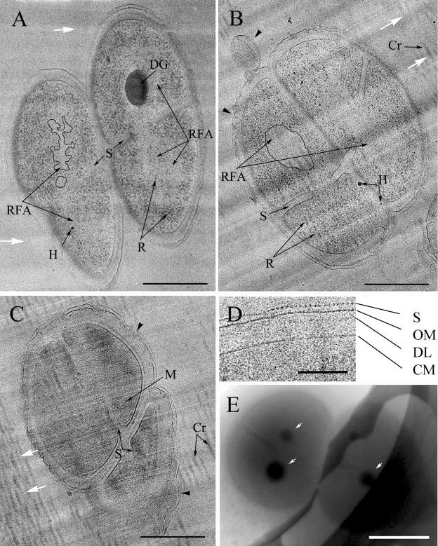
(A, B, C) Vitreous sections of typical D. radiodurans tetrads from exponentially growing (A), stationary-phase (B), and long-stationary-phase (C) cultures were imaged with high defocus (5 to 10 μm) to obtain strong contrast favorable for general morphology mapping. Knife marks (white arrows) and crevasses (Cr) are cutting artifacts. H, surface contamination with hexagonal ice. The cytoplasm of some adjacent cells in the tetrad is interconnected through incomplete septa (S). A large electron-dense granule (DG) is visible inside the exponentially growing cell. The small granules are ribosomes (R). In the central part of the exponentially growing and stationary-phase cells, ribosome-free areas (RFA) are seen and are outlined in one cell of the tetrad. Note the dispersed coralline shape of the ribosome-free areas in exponentially growing cells (A) and the compact roundish shape in stationary-phase cells (B). Some membranous structures (M) are the only distinguishable structures in the highly dense content of long-stationary-phase cells (C) in these imaging conditions. Irregularities in the structure of the outer part of the cell envelope (black arrowheads) are often observed in vitreous sections. (D) Magnified fragment of the cell envelope. The cytoplasmic membrane (CM), the diffuse dense layer (DL), the outer membrane (OM), and the outermost periodic S-layer (S) of the cell wall are distinguishable in vitreous sections. (E) Whole-mount exponentially growing tetrad plunge-frozen in culture medium. Note electron-dense granules (white arrows). Scale bars: A, B, and C, 500 nm; D, 100 nm; E, 1 μm.

