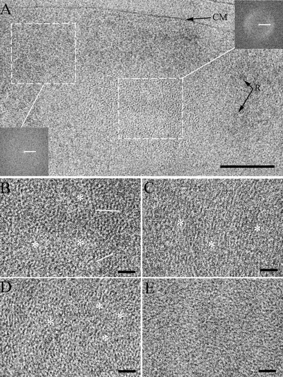
FIG. 3.
Fine structure of the nucleoid in stationary-phase cells. A, High-resolution view of stationary-phase cell (defocus, 2 μm). At this defocus the contrast of ribosomes (R) is faded, but fine structural details are visible in the nucleoid. Insets show Fourier transforms of the corresponding marked regions of the cell. CM, cytoplasmic membrane. B, C, D, and E, Different structural textures of the nucleoid. B, Dotted pattern. Regularly spaced arrays of dots are underlined in white. C, Stripy pattern. D and E, Intermediate case between dotted and stripy patterns which simultaneously contains dots and short and longer lines. The clusters of dots and lines are spaced by the regions without definable structure (asterisks). The aspects of the local order are less pronounced in some nucleoids (E). Scale bars: A, 100 nm; B, C, D, and E, 20 nm; Fourier transforms, (5 nm)−1.

