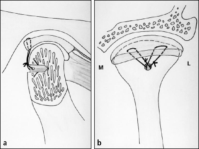
Figure 4.

(a) The cross-section of the condyle illustrates the Mitek anchor positioned beneath the posterior cortical bone, with the wings expanded, locking it in position. Usually, the anchor is inserted 8 to 10 mm below the condylar head and just lateral to the midsagittal plane. (b) A posterior view of the condyle shows the artificial ligaments (0-Ethibond sutures) secured to the posterior band of the repositioned articular disc. Two sutures (one posteromedial and the other posterolateral) are passed from the anchor to the disc in horizontal mattress fashion to stabilize the repositioned disc. M indicates medial; L, lateral.
