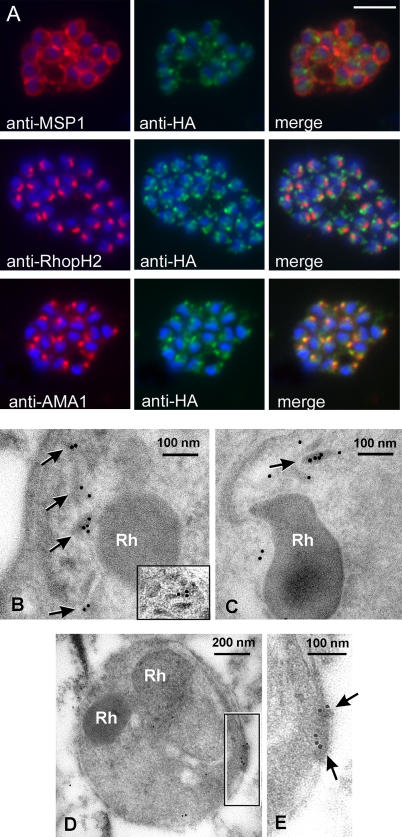
Figure 2. PfSUB2 Is a Microneme Protein.
(A) IFA images of schizonts of PfSUB2HA clone 2D dual-labelled with mAbs X509 (anti-MSP1), 61.3 (anti-RhopH2), or 4G2 (anti-AMA1), plus in each case mAb 3F10 (anti-HA) The anti-HA signal co-localised only with the anti-AMA1 signal. Identical results were obtained with the uncloned transgenic PfSUB2HA line, and/or when anti-RAP2 mAb H5 was used as the rhoptry marker instead of mAb 61.3 (not shown). Parasite nuclei are stained throughout with DAPI (blue). Scale bar represents 2 μm.
(B and C) Electron micrographs showing immuno-gold labelling of micronemes within late-stage schizonts of PfSUB2HA clone 2D using: (B) anti-HA mAb 3F10, detecting epitope-tagged PfSUB2; and (C) a polyclonal antibody specific for PfAMA1. The inset in (B) shows another example of micronemal staining with mAb 3F10 from another schizont. Rh, rhoptry.
(D and E) Posterior labelling of with anti-HA mAb 3F10 in a free merozoite of PfSUB2HA clone 2D. Arrows indicate immuno-gold labelling. Rh, rhoptry.

