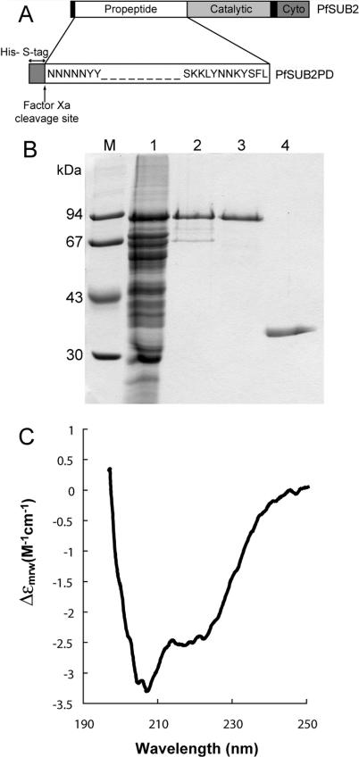
Figure 4. Recombinant PfSUB2 Propeptide Is Soluble and Structured.
(A) Schematic of the PfSUB2PD construct. Full-length PfSUB2 is represented with the secretory signal peptide and TMD indicated in black and the catalytic and cytoplasmic domains in shades of grey. PfSUB2PD possesses a cleavable 46-residue N-terminal fusion peptide containing a His6 and an S-tag, shown in dark grey.
(B) Coomassie-stained SDS PAGE gel showing stages in the purification of PfSUB2PD. Lane 1, Ni-NTA agarose eluate enriched in PfSUB2PD; lane 2, peak fraction from Superdex 200 gel filtration of previous lane in 8 M urea; lane 3, peak fraction from the final RP-HPLC step; lane 4, purified PfSUB1 propeptide (PfSUB1PD). Lane M contains molecular mass markers, the sizes of which are indicated.
(C) Far-ultraviolet CD spectrum of purified refolded PfSUB2PD. Deconvolution of the spectrum obtained calculated secondary structure values of 24% α-helix, 23% β-sheet, 21% reverse turn, and 32% random coil.

