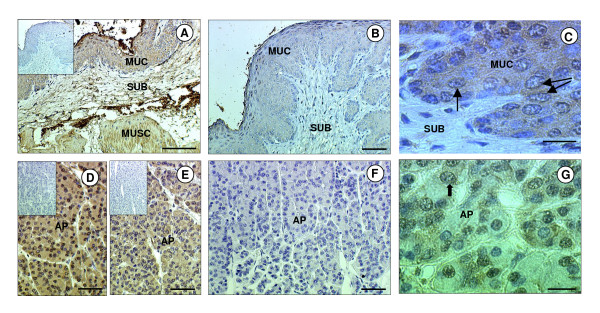
Figure 1.
Immunohistochemical localisation of ghrelin and GHSR-1a in the sheep stomach and pituitary gland. Photomicrographs of sheep stomach (abomasum) and anterior pituitary (AP) sections immunostained with antibodies against either ghrelin or GHSR-1a. (A) Stomach section showing positive staining for ghrelin (brown) in tunica mucosa (MUC), tunica submucosa (SUB) and tunica muscularis (MUSC). (B) Positive immunostaining in the stomach was abolished when the antiserum was pre-incubated with the immunising peptide (ghrelin). (C) High magnification micrograph of MUC showing the cytoplasmic and perinuclear (arrow) nature of the immunostaining for ghrelin. (D) AP section showing positive staining for ghrelin in most cells. (E) AP section showing positive staining for GHSR-1a in most cells. (F) Positive immunostaining for GHSR-1a in the AP was abolished when the antiserum was pre-incubated with the immunising peptide. (G) High magnification micrograph of the AP showing the cytoplasmic and perinuclear (arrows) nature of the immunostaining for GHSR-1a. The scale bar of A represents 100 μm, for B, D, E, F they represent 50 μm, and for C, G they represent 20 μm. The insert is the negative control.

