Figure 8.
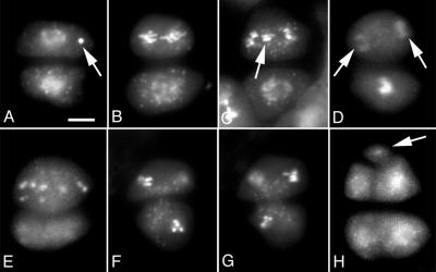
Asynchronous M-phase during meiosis II in tam. A, Dyad contained a chromosome fragment (arrow). B, Dyad with one cell at anaphase (top) and the other cell at prophase. The chromosomes in the top cell were less condensed than is typical for anaphase. C, Dyad with a phenotype similar to that shown in B. The top cells contained lagging chromosomes. D, One cell has completed nuclear division (arrows) and the other cell was still at late prophase or early metaphase stage. E, An abnormal tetrad with three cells. The larger cell was at anaphase and contained scattered chromosomes. The two smaller cells at bottom are out of focus. F and G, Two focal planes of the same dyad, showing that both cells were at telophase. H, An abnormal tetrad with five cells. DAPI staining was used. Scale bar, 5 μm.

