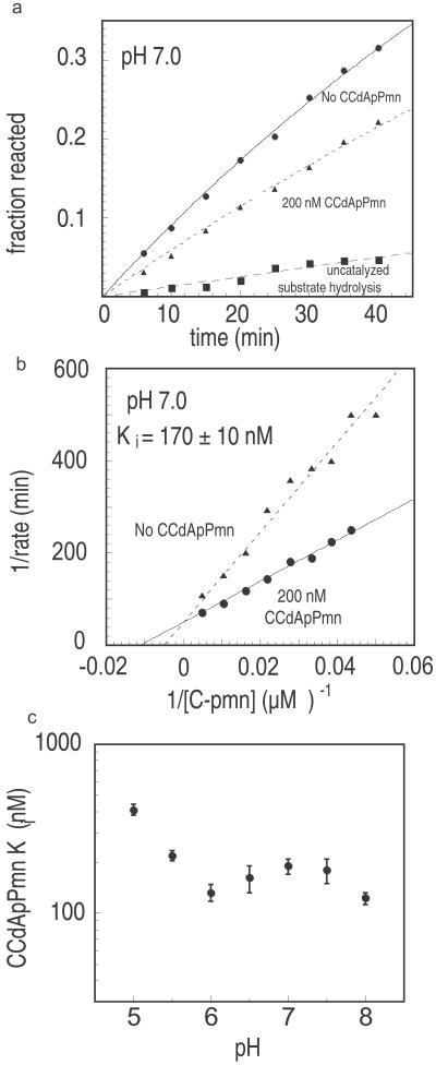
Figure 3.
pH dependence of CCdApPmn binding based on inhibition of the modified fragment assay. (a) Peptidyl transfer fragment assay inhibition by CCdApPmn at pH 7.0. Reactions contain 62 μM CPmn in the presence (200 nM) or absence of CCdApPmn. Also shown is the extent of background hydrolysis of the radiolabeled substrate CCA-pcb in the absence of CPmn. (b) Lineweaver–Burke plot of inhibition of the modified fragment assay at pH 7.0 in the presence or absence of CCdApPmn inhibitor. The concentrations of CPmn were 20, 23, 26, 30, 36, 46, 62, 95, or 200 μM. The Ki value (125 ± 10 nM) was calculated by using Eq. 2. (c) The pH dependence of Ki for CCdApPmn. Each point represents an average of two Lineweaver–Burke plots with two sets of nine CPmn concentrations each. Standard deviations are indicated by error bars.

