Figure 4.
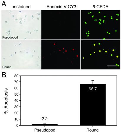
Evaluation of apoptotic activity in selected pseudopod and round gonocyte subpopulations by annexin V staining. (A) Selected pseudopod (Upper) and round (Lower) gonocytes were double-stained with CY3-labeled annexin V (red) and 6-CFDA (green) to identify apoptotic and viable cells, respectively. Phase-contrast microscopic images show homogeneous populations of pseudopod (A Upper Left) and round (A Lower Left) gonocytes. Pseudopod gonocytes rarely exhibit annexin V staining (A Upper Center), but many round gonocytes bind annexin V (A Lower Center), indicating that they are dying. Staining with 6-CFDA demonstrates that the majority of selected pseudopod (99%, A Upper Right) and round (92% A Lower Right) are viable. Overlapping red and green produces yellow fluorescence in most round gonocytes (A Lower Right), demonstrating that the annexin V staining can be attributed to apoptosis (a regulated event) rather than necrosis. The fluorescence results (A Center and Right) were obtained by using an epifluorescent microscope and Leitz N2 (Center, red: excitation, 530–560 nm; emission, >580 nm) and H3 (Right, green + red: excitation, 420–490 nm; emission, >515 nm) filter cubes. (Bar = 100 μm.) (B) Quantitative analysis of annexin V staining results indicate that only 2.2 ± 0.9% of viable pseudopod gonocytes are undergoing apoptosis compared with 66.7 ± 5.8% of viable round gonocytes. Necrotic cells were excluded from this analysis. Values (means ± SEM) were from eight replicate experiments encompassing 0–4 dpp. An average of 42.6 ± 4.0 cells was evaluated for each replicate.

