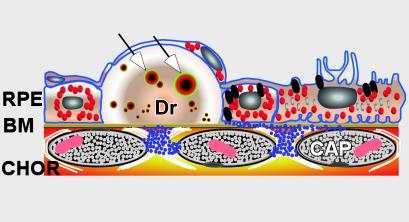
Figure 1.
Diagram depicting the anatomical relationships among the retinal pigmented epithelium (RPE), Bruch's membrane (BM), and drusen (Dr). Vesicles containing amyloid β that likely represent primary sites of complement activation are identified within drusen (arrows). CHOR, choroidal vasculature; CAP, capilllary.

