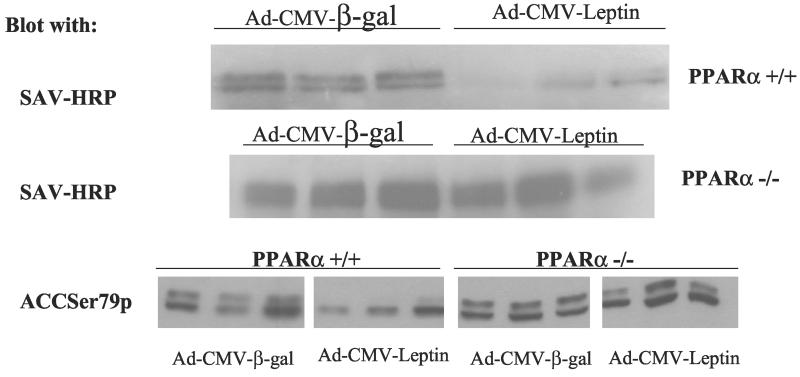
Figure 5.
ACC content in mouse liver. Shown are streptavidin–horseradish peroxidase blots (Top and Middle) of extracts of mouse adipose (in triplicate) prepared from PPAR+/+ and PPAR−/− mice 7 days after the i.v. administration of AdCMV-leptin or AdCMV-β-gal as a control. Visible are the two known ACC isoforms, ACCβ (280 kDa) and ACCα (265 kDa). (Bottom) The same samples have been blotted with an antibody that specifically recognizes Ser79p, the regulatory phosphorylation site of ACC.

