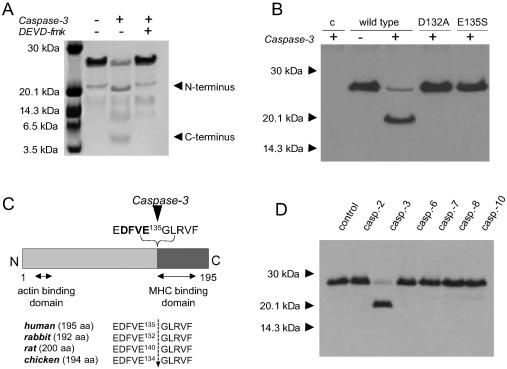
Figure 2.
Determination of the cleavage site of vMLC1 and its specificity for caspase-3. (A) Cleavage of purified human vMLC1 (10 μg) by recombinant active caspase-3 (20 ng/μl, 1 hr, 37°C), analyzed by SDS/16.5% PAGE and Coomassie blue staining. (B) Immunoblot analysis of wild-type vMLC1, vMLC1mD132A, and vMLC1mE135S proteins expressed in COS-7 cells, after incubation of the cell extracts with recombinant active caspase-3 (20 ng/μl, 1 hr, 37°C). The first line (c) shows extracts from COS-7 cells transfected with control vector plasmid. Mouse monoclonal antibody directed against the residues 1 to 8 (clone F109.17A5) was used for immunodetection. (C) A diagram showing the caspase-3 cleavage site at the C-terminal side of E135 of human, rabbit, rat, and chicken vMLC1. (D) Immunoblot analysis of vMLC1 cleavage in protein extracts from rabbit left ventricle, incubated with different human recombinant active caspases (25 ng/μl) for 1 hr.

