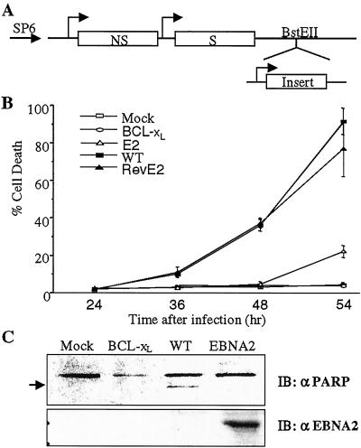
Figure 1.
Inhibition of SV-induced cell death by EBNA2. (A) Diagram of the SV vector. SP6, heterologous SP6 promoter; NS, nonstructural genes; S, structural genes. (B) NIH 3T3 cells were mock infected or infected at a moi = 5 with SV-wt, SV-BCL-xL, SV-EBNA2 (E2), or SV-revEBNA2 (revE2). Cell viability was determined by trypan blue exclusion. The data shown are the mean of three assays with the standard deviation provided. (C) NIH 3T3 cells were infected with virus at a moi = 5, and harvested at 48 hr after infection. Intact PARP and the 85-kDa cleaved product (arrowed) were detected by Western blotting. The same blot was stripped and reprobed with anti-EBNA2 antibody (Lower).

