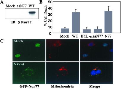
Figure 2.
Nur77 is a mediator of SV-induced apoptosis. (A) NIH 3T3 cells (2 × 10⁷) were infected with either SV-wt or SV-antisenseNur77 (asN77) for 4 hr. Cell lysates were immunoprecipitated with anti-Nur77 antibody and the immunoprecipitates were analyzed by immunoblotting with anti-Nur77 antibody. (B) NIH 3T3 cells were mock infected or infected at a moi = 5 with SV-wt, SV-BCL-xL, SV-Nur77 (N77), or SV-antisenseNur77 (asN77). Cell death was measured 48 hr after infection by using an LDH release assay. The data shown are the mean of three assays with the standard deviation provided. (C) NIH 3T3 cells were transfected with GFP-Nur77 and 12 hr later the cells were either mock infected (Upper) or infected with SV-wt (Lower). After 24 hr, cells were fixed, and stained with DAPI (nuclei; blue) and MitoTracker-Red (mitochondria; red). GFP-Nur77 (green) was nuclear in mock-infected cells (Upper) but colocalized with mitochondria in SV-infected cells.

