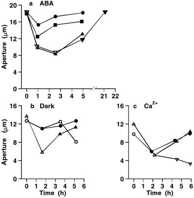
Figure 6.
3,4 DP prevents stomatal closure induced by ABA, dark, and Ca2+. The closing signal was applied immediately after the first aperture measurements, and changes in aperture followed with time. In each curve, white and black symbols show measurements in the absence and presence of 3,4 DP. (a) 10 μM ABA, 40 μM 3,4 DP. Each curve shows the mean of two replicate strips. (b) Dark, 25 μM 3,4 DP. Four strips were closed in the dark in the absence of inhibitor, and 3,4 DP was added after 1.6 h (▵, ▴). Four strips were treated in the dark in the presence of 3,4 DP; 3,4 DP was then removed from two strips (○), and two were left in 3,4 DP (●). (c) 10 mM external Ca2+, 25 μM 3,4 DP. Six replicate strips were transferred to 3,4 DP from zero time (○, ●). Of the six control strips closed in the absence of 3,4 DP (▵), four were treated with 3,4 DP after 2.2 h (▴), and two remained in the absence of inhibitor (▿). SEs (not shown) were in the range of 3% to 10%.

