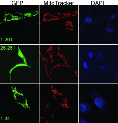
Fig 2.
Subcellular localization of N-terminal fragments of hTop3α fused to EGFP. Fluorescence images of HeLa cells, transiently transfected with the respective construct indicated in the leftmost image of each row, are depicted. The three images of each row show the patterns of the green EGFP fluorescence (Left), the mitochondria-specific dye MitoTracker Red (Center), and the DNA-specific reagent 4,6-diamino-2-phenylindole (DAPI) that prominently stains the nuclei (Right).

