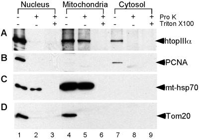
Fig 4.
Presence of endogenous human DNA topoisomerase IIIα in both the nucleus and mitochondria. HeLa cells were collected and separated into nuclear (lanes 1–3), mitochondrial (lanes 4–6), and cytosolic fractions (lanes 7–9), as described in Materials and Methods. For each subcellular fraction, the samples were incubated for 30 min on ice in the absence of proteinase K (lanes 1, 4, and 7), in the presence of proteinase K (lanes 2, 5, and 8), or in the presence of both proteinase K and Triton X-100 (lanes 3, 6, and 9), as indicated on top of the figure. (The plus sign indicates the presence and the minus sign the absence of the reagent specified in the rightmost column; pro K, proteinase K.) The treated samples were then subjected to SDS/PAGE and the resolved protein bands were immunoblotted by using antibodies against human Top3α (A), PCNA (B), mt-hsp70 (C), and Tom20 (D). In each case, only one prominent band was detected, and its apparent molecular mass agreed with that expected of the target of the antibodies used (marked in the right-hand margin).

