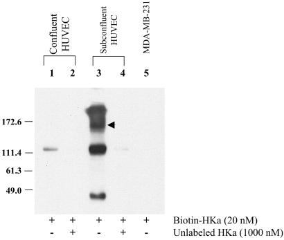
Fig 7.
Cross-linking of HKa to endothelial cell surface proteins. Biotin-HKa was incubated with confluent (lanes 1 and 2) or proliferating (lanes 3 and 4) endothelial cells in the absence (lanes 1 and 3) or presence (lanes 2 and 4) of a 20-fold molar excess of unlabeled HKa before cross-linking by using bis(sulfosuccinimidyl) suberate. Biotin-HKa was incubated also with MDA-MB-231 breast carcinoma cells under identical conditions (lane 5). Detergent extracts were separated by SDS/PAGE and then transferred to poly(vinylidene difluoride) and detected by chemiluminescence. The arrowhead denotes a prominent band of ≈140–150 kDa, the expected size of an HKa-tropomyosin complex. HUVEC, human umbilical vein endothelial cells.

