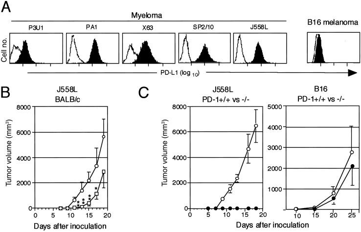
Fig 4.
Inhibition of the tumorigenesis of myeloma cells endogenously expressing PD-L1 in the normal syngeneic mice treated with anti-PD-L1 Ab or in the PD-1-deficient mice. (A) Expression of endogenous PD-L1 in myeloma and B16 melanoma cells was examined with a flow cytometry. (B) J558L myeloma cells (2.5 × 10⁵) were inoculated s.c. into the syngeneic BALB/c mice (nine mice per group) followed by the injection with normal rat IgG (○) or anti-PD-L1 mAb (□) (0.1 mg per mouse) on days 1, 3, 5, and 7. The mean tumor volumes and SE of nine mice are indicated. **, P < 0.01; *, P < 0.1 (by Student's t test). (C) J558L (Left, 2.5 × 10⁵) or B16 (Right, 1 × 10⁶) cells were inoculated s.c. into PD-1+/+ (○) and −/− (•) mice of BALB/c or B6 background, respectively. The mean tumor volumes and SE of four (the former) and 10 (the latter) mice are indicated.

