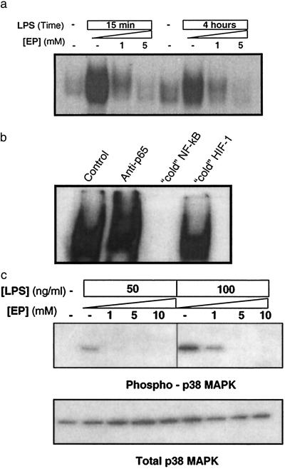
Fig 4.
EP prevents LPS-induced activation of NF-κB and p38 MAPK pathways in macrophage cultures. (a) RAW264.7 cells were stimulated with 50 ng/ml or 100 ng/ml LPS in the presence of the indicated concentrations of EP. NF-κB activation was analyzed at 15 min and 4 h by electrophoretic mobility-shift assay by using a previously described 32P-labeled NF-κB probe. EP suppressed NF-κB activation in a concentration-dependent fashion. The results shown are representative of an experiment that was repeated three times. (b) Nuclear extracts of LPS-stimulated macrophages were incubated with antibody against p65-Rel (“anti-p65”) to induce specific supershift of NF-κB complex or 100-fold molar excess of unlabeled (cold) NF-κB or HIF-1 irrelevant duplex DNA probe for competition analysis. The results shown are representative of an experiment that was repeated three times. (c) RAW264.7 cells were stimulated with LPS (50 or 100 ng/ml) in the presence of the indicated concentration of EP. The phosphorylation of p38 MAPK was analyzed by Western blot by using antibodies against phosphorylated (Thr-180/Tyr-182) p38 MAPK, in accordance with the manufacturer's instructions (9210; New England Biolabs). EP prevented the phosphorylation of p38 MAPK in a concentration-dependent fashion (Upper), without affecting the intracellular level of total p38 MAPK (Lower). The results shown are representative of an experiment that was repeated three times.

