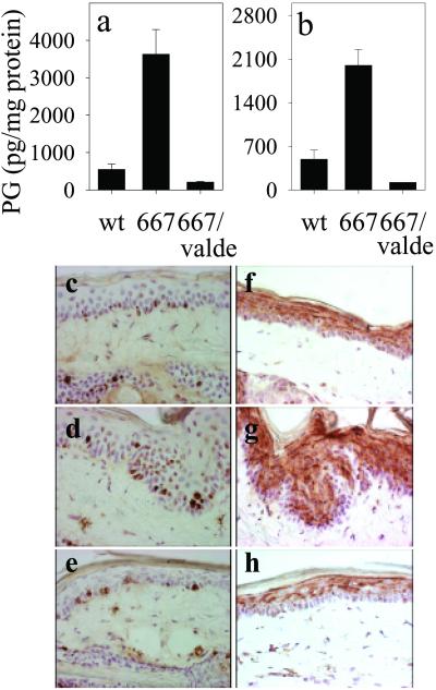
Fig 2.
Suppression of the COX-2 transgene-induced PG levels and phenotype by COX-2 inhibition. The levels (mean ± SEM) of PGE2 (a) and PGF2a (b) were determined for tail skin of wt NMRI (n = 7), K5.COX-2/667+/− transgenic mice fed with a control diet (667, n = 8), and K5.COX-2/667+/− transgenic mice receiving a valdecoxib-containing (valde) diet (667/valdecoxib, n = 3); P values for PGE2 and PGF2α were ≤0.0001. The histology and localization of Ki67-positive keratinocytes in the interfollicular epidermis of tail-skin sections are shown for wt (c), K5.COX-2/667+/− transgenic (d), and valdecoxib-treated K5.COX-2/667+/− transgenic mice (e). K10 staining in tail-skin sections is shown for wt (f), K5.COX-2/667+/− transgenic (g), and valdecoxib-treated K5.COX-2/667+/− transgenic mice (h). [Magnification, ×400 (c–h).]

