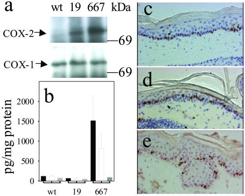
Fig 3.
COX isozyme expression, PG levels, and skin morphology in age-matched transgenic as compared with wt mice. (a) COX isozyme expression in epidermis from wt mice and the transgenic lines K5.COX-2/19+/− (19) and K5.COX-2/667+/− (667). (b) Epidermal PG levels (mean ± SEM; PGE2, black bars; PGF2α, white bars; 6-keto-PGF1α, gray bars) in wt mice (n = 5) and the transgenic mice K5.COX-2/19+/− (19, n = 5) and K5.COX-2/667+/− (667, n = 3); P values for PGE2 and PGF2α (wt vs. K5.COX-2/667+/−) were ≤0.036. Epidermal histology and localization of Ki67-positive keratinocytes are shown for skin sections from wt mice (c) and the transgenic mice of K5.COX-2/19+/− (d) and K5.COX-2/667+/− (e). [Magnification: ×400.]

