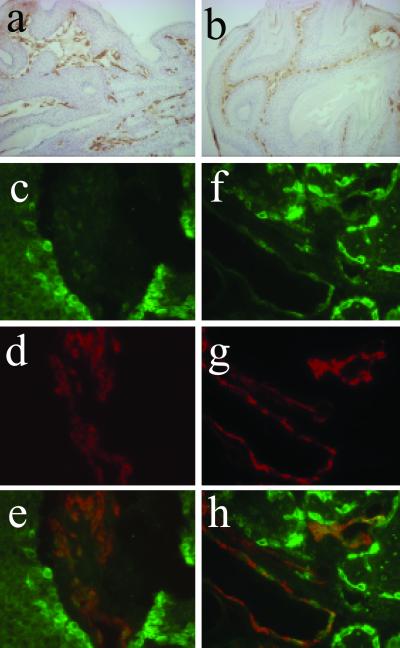
Fig 5.
Localization of COX-2 protein in the tumor vasculature. Tumor vasculature is visualized in a papilloma derived from a DMBA-initiated K5.COX-2/667+/− transgenic mouse (a) and a DMBA/PMA-treated wt mouse (b) by immunostaining for the endothelial cell marker CD31. A double-immunofluorescence analysis was performed for a papilloma (c–e) and a sebaceous gland adenoma (f–h) from DMBA-initiated K5.COX-2/667+/− transgenics by using anti-COX-2 and anti-CD31 antisera revealing that COX-2 (c and f, green) and CD31 (d and g, red) colocalize in tumor vessels (e and h, yellow). COX-2 expression is visible also in basal cells of papillomas and sebaceous gland adenomas (c and f). [Magnification: a and b, ×160; c–h, ×630.]

