Figure 3.
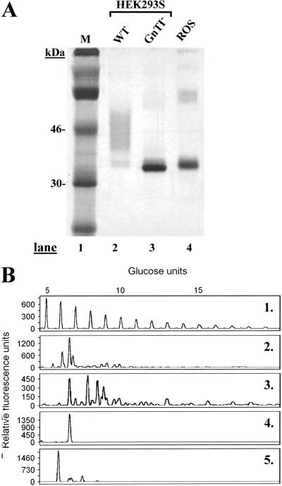
Characterization of rhodopsin and rhodopsin N-glycans prepared from HEK293S and HEK293S-GnTI− stable cell lines. The cell lines were induced for 2 days by using tetracycline and sodium butyrate. (A) Rhodopsin was purified and examined by SDS/PAGE (10%) followed by silver stain. Lane 1, M (molecular weight standards); lane 2, rhodopsin from inducible HEK293S; lane 3, rhodopsin from HEK293S GnTI−, and lane 4, rhodopsin from ROS. (B) Characterization of rhodopsin N-glycan composition. Profile 1, mobilities of molecular weight standards (malto-oligosaccharides) used for calibration. Profiles 2 and 3, N-glycans from rhodopsin samples prepared from inducible HEK293S cell line (before and after treatment with sialidase, respectively); profile 4, rhodopsin from HEK293S GnTI−; and profile 5, ROS rhodopsin.

