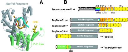
Figure 1.
Schematic representation of chimeric polymerases. (A) Domain organization of TaqDNA polymerase in which helices are represented by cylinders and β-strands by arrows. This structure has been modeled by using two available x-ray structures of Taq polymerase (in “open” and “closed” conformations; for details, see Text, which is published as supporting information on the PNAS web site). The polymerase and inactive 3′-5′ exonuclease domains are colored gray, and the 5′-3-exonuclease domain is colored green. Several amino-terminal and carboxyl-terminal amino acids are colored magenta and red, respectively. The only HhH motif in the 5′-3′ exonuclease domain is colored gold. DNA strands are colored cyan and orange. (B) Cartoon illustration of chimeric constructs. HhH repeats of Topo V are colored yellow (H–L), orange-yellow gradient (E–G), orange (C and D), and rainbow (A and B). Arrows indicate cleavage positions that result in C1–C3 domains (in case of Topo V) and the Stoffel fragment (in case of Taq polymerase).

