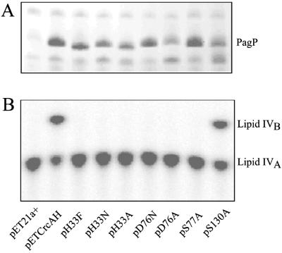
Figure 2.
Expression and activity of PagP-His6 and its site-directed mutant derivatives. (A) Membrane proteins were analyzed by SDS/PAGE and stained with Coomassie blue dye (10). The band corresponding to fully folded PagP is indicated. Note that unfolded PagP migrates more rapidly and did not accumulate for any of the mutants. (B) Palmitoyl transferase reactions were performed with induced membranes from the host strain BL21(DE3)pLysE at 100 ng/ml using 32P-lipid IVA as the acyl-acceptor, and the production of the palmitoylated metabolite lipid IVB was detected by TLC (10). The compounds on the plate were visualized by overnight exposure to a PhosphorImager (Molecular Dynamics) screen. pET21a+-transformed cells were used as a negative control, whereas pETCrcAH-transformed cells expressing wild-type PagP served as a positive control.

