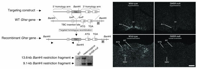
Figure 1.
Gene targeting of the Ghsr locus. Upper left panel: Schematic diagram of the derivation of GHSR-null mice by homologous recombination. Lower left panel: Southern blot analysis of genomic DNA extracted from representative progeny of mating animals heterozygous for the recombinant Ghsr allele. Right panels: Representative dark-field photomicrographs of in situ hybridization histochemistry experiments performed on mouse brains using a mouse GHSR–specific riboprobe. Ghsr mRNA expression is evidenced by the white-appearing silver granules. 1–3, Ghsr exons 1–3; 3V, third ventricle; AP, area postrema; Arc, arcuate nucleus; DMV, dorsal motor nucleus of the vagus; NTS, nucleus of the solitary tract; P, Southern probe. Scale bar: 200 μm (applies to all 4 panels).

